




Xiaogan Kuohai Medical Technology Co., Ltd. is one of the most reliable manufacturers and suppliers of rotary microtome in China, featured by quality products and good price. If you're going to wholesale advanced rotary microtome made in China, welcome to get quotation from our factory. Also, customized service and 1 year warranty are available.
Company Profile
Xiaogan Kuohai Medical Technology Co.,Ltd. is a rare domestic pathology instrument manufacturing company with a widespread user base throughout China. It is also one of the few companies in the domestic and international industry capable of researching, developing, and producing a complete set of pathology instruments and consumables. In 2014, the company established a modern production and processing base.
Why Choose Us
Rich Experience
Xiaogan Kuohai Medical Technology Co., Ltd. is a rare domestic pathology instrument manufacturing company with a widespread user base throughout China.
High Quality
The product range covers industries such as medical instruments, biotechnology, electronic technology, high-end building materials, and decoration engineering.
Technical Support
Our team of experts is available to assist with troubleshooting, answer technical inquiries, and provide guidance.
Customer Service
We prioritize open communication to address our clients' specific requirements and deliver personalized solutions.

The tool holder selects special materials through special surface treatment technology, so that the slices are smooth and continuous, and the wax pieces will not adhere to the tool holder, and good slicing effect and slice quality are obtained.

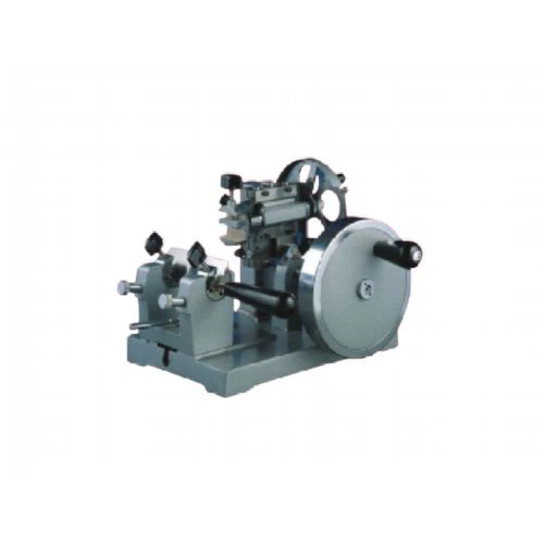
The pathological rotary slicer is a third-generation improved model based on the original KH-Q300A slicer, which has greatly improved in accuracy and appearance.

Equipped with intelligent key function (can be operated blindly without looking at the screen), the main functions of the slicer can operate normally when there is no display on the screen and the touch function is damaged;

New cross roller guide rail, durable lubrication micro-feed mechanism, no need to add lubricants maintenance;

4.3-inch color touch screen, can display the number of slices and the total thickness of slices.

The pathological rotary microtome is a third-generation improved model based on the original KH-Q300A paraffin microtome, which has greatly improved in accuracy and appearance. It has become a cost-effective, practical and complete slicer.
A rotary microtome is a laboratory instrument used to prepare thin sections of biological specimens for microscopic examination. The microtome uses a rotating blade to cut the specimen into thin slices, usually ranging from a few micrometers to several hundred micrometers in thickness. The slices are then mounted on slides and stained for observation under a microscope.
Rotary microtomes are commonly used in fields such as histology, anatomy, and pathology to prepare samples for light and electron microscopy. They are also used in material science and other fields where precise thin sectioning of materials is required.
A rotary microtome consists of a rotating blade, a sample holder, and a stage for positioning the specimen. The sample holder holds the specimen in place while the blade rotates and cuts the slices. The thickness of the slices can be adjusted by changing the distance between the blade and the specimen or by changing the blade itself.
Benefits of Rotary Microtome
Micrometer-Driven Specimen Advance
Rotary microtomes are equipped with a micrometer-driven specimen advance mechanism that allows precise control over section thickness. This feature enables researchers to obtain consistent and reproducible sections, crucial for accurate analysis.
Adjustable Knife Angle and Speed
Rotary microtomes provide the flexibility to adjust the knife angle and speed, allowing researchers to optimize sectioning parameters based on tissue type and characteristics. This adaptability ensures optimal cutting performance and minimizes tissue damage.
Section Waste Tray and Collection System
Modern rotary microtomes are designed with section waste trays and collection systems, facilitating easy retrieval and organization of tissue sections. This feature enhances workflow efficiency and reduces the risk of section loss or contamination.
Ergonomic Design
Otary microtomes have undergone ergonomic improvements, offering user-friendly interfaces, adjustable handwheels, and comfortable hand rests. These design enhancements prioritize user comfort, reducing fatigue during extended sectioning sessions.
Type of Rotary Microtome
Sled microtomes
Sled microtomes place the sample in a fixed holder or shuttle which moves back and forth across a knife or blade. Typically, the sled rests upon a linear bearing and permits angular adjustment. Often, sled microtomes are used with biological materials and samples that are embedded in paraffin.
Vibrating microtomes
Vibrating microtomes use a vibrating blade which facilitates the sample cutting. This microtome uses less pressure than would be required with a stationary blade and is often used for difficult biological samples such as nervous tissues, brain and spinal cord. The stability of the instrument reduces the mechanical forces on the tissue sample to ensure the retention of high-quality viable cells.
Infrared laser microtomes
Infrared laser microtomes are used to achieve contact-free slicing. To provide precise control, only small pulses of infrared (IR) energy are imparted into the sample. Laser microtomes offer several advantages including non-contact cutting, no sample preparation for native tissue, no decalcification of hard tissue, and a cut with precision of 1µm. They are generally too slow for routine pathology applications.

Factors To Consider While Buying A Rotary Microtome
Good Section Quality
It is vital to ensure the quality of the sections when working on a research project or diagnosing a disease. A quality section means precision and stability of tissues and quality of different components of microtome, such as
Feeding-mechanism precision
When the specimen is orientated precisely with a zero-point reference, the block can be aligned quickly and re-cut as needed. Moreover, the electric cooling specimen device in microtomes helps you to obtain uniform sections. However, this feature is optional and isn't present in all rotary microtomes.
Safety Features in Microtomes
An object head that is adjustable depending on the clamp's size and weight. It prevents accidental drops onto the blade during sectioning.
With the handwheel safety locks, you can exchange blades and specimens easily and adjust specimen orientation without the object head moving.
Blade ejectors and magnetic brushes make it easy to remove blades from blade holders without touching them.
Microtomes equipped with safety guards enable you to cover the blade when they are not in use. As a result, the blades of the microtome can be cleaned and replaced without touching them.
Ensure the coarse feed wheel is as close to the user as possible so that ergonomic comfort extends for a long time when rock trimming.
Type of Operation
Although manual machines are less expensive than automated ones, automated rotary microtomes have many advantages in terms of ergonomics and safety features:
Using hand wheels repetitively for many hours can lead to discomfort and increase the risk of work-related musculoskeletal disorders (WMSDs). This is avoidable by using an automated rotary microtome which eliminates the need of using the handwheel.
A built-in safety mechanism to prevent uncontrolled motorized sectioning. For example, sectioning should have two separate buttons to begin the process, this is available in most brands.
During an emergency, the motor is immediately stopped by an emergency stop button located on the instrument housing or foot switch.
How to Choose the Right Rotary Microtome
Sectioning Precision and Thickness
The primary function of a rotary microtome is to produce thin, uniform tissue sections for microscopic analysis. Consider the sectioning precision and thickness range offered by different microtome models. Look for instruments that provide a wide range of section thickness settings to accommodate diverse tissue types and experimental requirements. Instruments with precise control mechanisms and fine adjustments ensure reproducibility and consistency in sectioning.
Sample Size and Compatibility
Evaluate the range of sample sizes that the rotary microtome can accommodate. Different microtome models are designed to handle varying sample dimensions, from small biopsies to larger tissue blocks. Choose a microtome with adjustable specimen clamps or specimen orientation features to enhance flexibility in sample positioning. Compatibility with a variety of specimen molds and chucks allows for efficient sectioning of different tissue shapes and sizes.
Ergonomics and User-Friendly Design
The ergonomic design of a rotary microtome contributes significantly to user comfort and operational efficiency. Look for features such as a user-friendly control panel, easy-to-operate handwheel, and comfortable hand rests. Some advanced models offer touch-screen interfaces and programmable settings for enhanced user experience. Ergonomic considerations become particularly important for laboratories with high sample throughput or extended sectioning sessions.
Routine histological technique - In histology, tissues are normally hardened by replacing water with alcohol. Once the tissue is hardened, a microtome is used to cut and produce sections of varying thicknesses.
This technique is particularly useful in cases where the goal is to analyze the tissue within the shortest period of time possible. As compared to some of the other techniques, this technique makes it possible to study cells and other parts of the specimen in their native state (or near their native state).
Botanical microtechnique - Botanical Microtechnique is part of botanical histology used to study plant specimens at the microscopic level. For this reason, a Rotary Microtome is used to produce sufficiently thin sections to be analyzed under the microscope.
As compared to animal specimens, plant specimens are harder which necessitates the use of diamond knives (microtome knives). These knives are specifically used to cut a range of harder material including bone and plants etc.
While a Rotary Microtome is used to cut plant specimens, it is worth noting that for the most part, these sections tend to be thicker compared to those of animal specimens.
Electron microscopy - In electron microscopy, very thin sections are required for analysis of the specimen. Commonly known as ultra-thin sections (as thin as 69nm in thickness), these sections are prepared using an ultramicrotome for transmission electron microscopy.
Automatic Microtomevs vibratome
A automatic microtome is a device used to cut very thin slices of tissue, called sections, for examination under a microscope. It typically uses a sharp, stationary blade to cut the tissue, and the thickness of the sections can be adjusted by changing the blade or adjusting the settings on the automatic microtome.
A vibratome is a device that uses a vibrating blade to cut tissue into sections. The vibration of the blade helps to reduce tissue tearing and produce cleaner, more accurate sections.
One key difference is the way in which the tissue is cut. A automatic microtome uses a stationary blade to slice the tissue, while a vibratome uses a vibrating blade. This can affect the quality and accuracy of the sections produced. Additionally, automatic microtomes are typically used to produce thinner sections, whereas vibratomes are better suited for thicker tissue samples.
There are also differences in the precision and accuracy of the sections produced by automatic microtomes and vibratomes. automatic microtomes are typically more precise, as the blade is stationary and can produce very thin, uniform sections. Vibratomes can produce sections that are slightly less precise, but are still suitable for many types of research and medical applications.
Ultimately, the choice between a automatic microtome and a vibratome depends on the specific requirements of the research or application, including the type and thickness of the tissue being studied, the desired thickness of the sections, and the precision and accuracy needed.
Both automatic microtomes and vibratomes are commonly used in research and medical laboratories to prepare tissue samples for microscopic examination. They are essentialtools for many types of scientific and medical research, including histology, cell biology, and pathology.
Major Rotary Microtome Parts For Day-to-day Operations
This is to reduce vibration during microtomy, in which stability is critical during sectioning to prevent undulations (washboarding) in the paraffin sections. Daily cleaning from paraffin debris and yearly preventive maintenance will keep a microtome cutting optimally for many years.
The main components of a rotatory microtome are described below. Although some microtomes have more bells and whistles, the standard microtome remains relatively simple to operate.
Microtome base plate or stage: A platform which has rails that secure the knife holder base.
Knife holder base: A part that anchors the knife holder to the microtome stage. The knife holder base can be moved toward or away from the block, but MUST be stationary and locked during microtomy.
Knife holder
This part is comprised of several components including the blade clamp that holds the blade, the knife tilt for adjusting the knife angle, and the face plate that guides that ribbons away from the blade and towards the operator.
Cassette clamp or block holder
Holds the paraffin block in place. Typically, the block moves up and down with each revolution while the blade is stationary. The block holder may have knobs that allow the user to manipulate the block face in various directions to bring the tissue in alignment with the blade.
Coarse handwheel
Moves the block holder either toward the knife or away from the knife.
Advancement handwheel
Turns in one direction and advances the block toward the knife at the specified microns. Most handwheels are equipped with a safety lock to prevent the wheel from releasing and having the block holder come down towards the blade while a block is inserted or removed. The safety lock should be used anytime the microtomist is not actively sectioning paraffin blocks.
Micron adjustment
Micron settings for section thickness can range from 1 to 60 microns on most microtomes.

Xiaogan Kuohai Medical Technology Co., Ltd. is a rare domestic pathology instrument manufacturing company with a widespread user base throughout China. It is also one of the few companies in the domestic and international industry capable of researching, developing, and producing a complete set of pathology instruments and consumables.
FAQ
Hot Tags: rotary microtome, China rotary microtome manufacturers, suppliers, factory, Semi automatic Microtome, Frozen Microtome, Manual Microtome, Tissue Microtome, Automatic Microtome, Microtome


