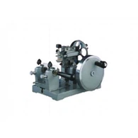


Xiaogan Kuohai Medical Technology Co., Ltd. is one of the most reliable manufacturers and suppliers of manual microtome in China, featured by quality products and good price. If you're going to wholesale advanced manual microtome made in China, welcome to get quotation from our factory. Also, customized service and 1 year warranty are available.
Xiaogan Kuohai Medical Technology Co.,Ltd
Xiaogan Kuohai Medical Technology Co.,Ltd. is a rare domestic pathology instrument manufacturing company with a widespread user base throughout China. "Kuohai Medical Technology" owns five subsidiaries, including Hubei Xiaogan Kuohai Medical Technology Co.,Ltd., Xiaogan Kuohai Medical Technology Co.,Ltd., Hubei Haishi Industrial Co.,Ltd., Xiaogan Ruifeng Electronic Technology Co.,Ltd., and Xiaogan Dinghang Decoration Engineering Co.,Ltd. The product range covers industries such as medical instruments, biotechnology, electronic technology, high-end building materials, and decoration engineering. The company is steadily moving towards industrial group development.
Why Choose Us
Rich Experience
Xiaogan Kuohai Medical Technology Co., Ltd. is a rare domestic pathology instrument manufacturing company with a widespread user base throughout China.
High Quality
The product range covers industries such as medical instruments, biotechnology, electronic technology, high-end building materials, and decoration engineering.
Technical Support
Our team of experts is available to assist with troubleshooting, answer technical inquiries, and provide guidance.
Customer Service
We prioritize open communication to address our clients' specific requirements and deliver personalized solutions.
Our Related Products

A fully automated microtome has both specimen feed and rotary motion of the handwheel automated, contributing to the ability to produce high-throughput sectioning. The mechanization of the instrument allows for automated setting and correction of feed movement and knife cutting. In addition, automation also provides consistency from user to user, so it can speed up time and provide reproducibility of sectioning. Many fully automatic microtomes allow for cutting a more comprehensive range of thicknesses, providing researchers flexibility when choosing sectioning materials such as paraffin or resin-based fillers.

A semi-automated microtome uses a specimen feed that is motorized, but the rotary motion of the handwheel is still done manually and offers a medium throughput. The semi-automated method works well for thick and hard sections, using a consistent force when cutting through a block. Automating the specimen feed provides the precision of a motorized specimen feeding that yields consistent, reproducible sections.

New cross roller guide rail, durable lubrication micro-feed mechanism, no need to add lubricants maintenance.
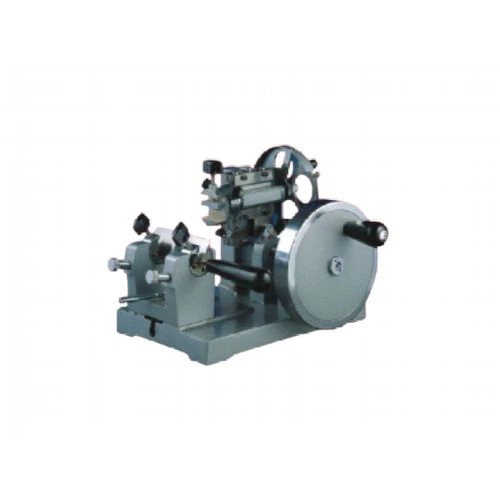
A microtome is a cutting tool used to produce skinny slices of material known as sections, with the process being termed micro sectioning. Important in science, microtomes are used in microscopy for the preparation of samples for observation under transmitted light or electron radiation.

Freezing microtome(cryostat) freezes the specimen to harden it so it can be sliced without ripping and destroying it. This microtome is designed for accurate and quick diagnosis in surgery and for the inspection of the quality of rubbers and others. It has a table clamping device.

A frozen microtome (sometimes used with a cryostat) is designed to freeze a specimen so that it can be hard enough to be accurately sliced by the microtome without destroying it. The freezing microtome can utilise temperatures typically ranging from -60 °C to 0 °C for freezing.

The rotary microtome is a manually operated cutting device for all types of work with paraffin and hard-cutting technology in the fields of biology, medicine, and industry. It is used for making microscopic preparations for subsequent illumination (e.g., human tissue).
What is Manual Microtome
Manual microtome is a rotary microtome specifically designed for manually cutting paraffin sections. It is ideal for routine use in clinical histology and histopathology, but is also capable of producing the finest cuts for research applications. One of its key features is a retraction mechanism that offers multiple advantages in the cutting process, including prolonging the lifespan of the blade. When operating the microtome in retraction mode, the entire block advances by the sum of the retraction value and the thickness of the chosen section upon a full rotation of the flywheel. Additionally, the microtome is equipped with a dual mechanical trimming device located at the bottom left, which significantly speeds up the roughing process. The unit features a casing made from a specialised solvent-resistant plastic that has no sharp edges, thereby reducing the risk of injury to operators.
Precision
Manual microtome allow for precise and consistent sectioning of tissue samples, ensuring that the thickness of each section is uniform and reproducible. This is critical for accurate diagnosis and research.
Versatility
Manual microtome can be used to prepare tissue samples from a wide range of sources, including human and animal tissues, plant tissues, and materials. This makes them a versatile tool for use in many fields of research.
Efficiency
Manual microtome are designed for rapid and efficient sectioning of tissue samples, allowing researchers and pathologists to process large numbers of samples in a relatively short amount of time.
High-quality sections
Manual microtome produce thin tissue sections with high clarity and minimal distortion, which is essential for accurate interpretation of tissue structure and morphology.
Flexibility
Manual microtome can be adjusted to produce tissue sections of varying thicknesses, allowing researchers and pathologists to tailor the sectioning process to their specific needs.
Application of Manual Microtome
Traditional Histology Technique
Tissues are fixed, dehydrated, cleared, and embedded in melted paraffin, which when cooled forms a solid block. The tissue is then cut in the microtome at thicknesses varying from 2 to 50 μm. From there the tissue can be mounted on a microscope slide, stained with appropriate aqueous dye(s) after removal of the paraffin, and examined using a light microscope.
Frozen section procedure
Water-rich tissues are hardened by freezing and cut in the frozen state with a freezing microtome or microtome-cryostat; sections are stained and examined with a light microscope. This technique is much faster than traditional histology (5 minutes vs 16 hours) and is used in conjunction with medical procedures to achieve a quick diagnosis. Cryosections can also be used in immunohistochemistry as freezing tissue stops degradation of tissue faster than using a fixative and does not alter or mask its chemical composition as much.
Electron Microscopy Technique
After embedding tissues in epoxy resin, a microtome equipped with a glass or gem grade diamond knife is used to cut very thin sections (typically 60 to 100 nanometer). Sections are stained with an aqueous solution of an appropriate heavy metal salt and examined with a transmission electron microscope. This instrument is often called an ultramicrotome. The ultramicrotome is also used with its glass knife or an industrial grade diamond knife to cut survey sections prior to thin sectioning. These survey sections are generally 0.5 to 1 μm thick and are mounted on a glass slide and stained to locate areas of interest under a light microscope prior to thin sectioning for the TEM. Thin sectioning for the TEM is often done with a gem quality diamond knife. Complementing traditional TEM techniques ultramicrotomes are increasingly found mounted inside an SEM chamber so the surface of the block face can be imaged and then removed with the microtome to uncover the next surface for imaging. This technique is called serial block-face scanning electron microscopy (SBFSEM).
Botanical Microtomy Technique
Hard materials like wood, bone and leather require a sledge microtome. These microtomes have heavier blades and cannot cut as thin as a regular microtome.
Spectroscopy (especially FTIR or infrared spectroscopy) Technique
Thin polymer sections are needed in order that the infra-red beam can penetrate the sample under examination. It is normal to cut samples to between 20 and 100 μm in thickness. For more detailed analysis of much smaller areas in a thin section, FTIR microscopy can be used for sample inspection.
How to Buying a Manual Microtome
Before buying a manual microtome for your facility, it's essential to understand the purpose of your experiments and the samples or specimens you will be working with.There is a range of manual microtomes with different knife profiles, sizes, shapes, and the ability to cut through soft to hard materials; thus, different experiments require different thicknesses of materials.
For example, electron microscopy requires sections as thin as 40-100 nm, while light microscopy requires a section of 0.5 μm or higher.
If working with soft, fragile, and fatty tissues, you should use general manual microtomes, such as rotary microtomes. They allow you to control the knife and obtain serial and uniform sample sections.
Saw microtome is suitable to cut through glass, ceramic, bone, and resin embedded specimens. Materials as thick as 20µm can be cut using this microtome.
Microtome allows you to cut sections of plants and animals in blocks or batches. It's simple, easy, and affordable; however, its lightweight structure makes it difficult to prepare thin sections.
Vibrating microtomes are preferred while developing slides for fresh animals and plant materials.
A sledge microtome is preferable when working with a larger specimen (like the whole brain) or a heavier specimen (like bone and teeth). It's compatible with paraffin-embedded samples and celloidin-embedded blocks.
The thickness of the sections of your samples is important for microscopy quantification. You can't obtain clearer details using thicker samples; however, thinner samples might be ruined before being used in the experiment. Thus, choose the microtome that can provide sections that suit your application.
Another factor to consider is deciding what kind of knife you want to adjust in your tool. Different knives are designed to serve different purposes. For example, fixed knives made of steel are suitable for cutting paraffin-wax embedded tissues, while those made of glass are perfect for resin-embedded tissues.
Fixed knives are also built of tungsten carbide, non-corrosive metal, diamond, and sapphire and are cheaper than disposable knives.
The disposable blades are made of polymer, Teflon, titanium nitride (TiN), or ceramic and are suitable to prepare thin sections of any sample, except harder specimens.
The knives used in microtomes come in different forms. And based on their structure they are classified as:
● Biconcave: It's extremely sharp and has two hollow ground surfaces. It's used with a rocking microtome to section celloidin embedded materials.
● Strongly-plano concave: It is sharp and has one hollow grounded side and one straight side to cut through soft, embedded materials.
● Plano concave: It has a different degree of concavity and is primarily used to cut paraffin and nitrocellulose embedded tissues.
● Wedge: It can be used with any microtome to cut all kinds of sections though initially designed to cut frozen samples.
● Tool edge shaped: It's used to cut hard specimens, such as bone and samples embedded either in paraffin or synthetic resin. Though less sharp, it's more stable than other blades.
Importance of Manual Microtome
In the everyday routine inside a laboratory, microscopic substances and samples are very common. Most of the time it is necessary to cut some of the samples in extremely thin slices of material, known as sections. In order to prepare a sample and to make those types of cuts, you must have a manual microtome, a very essential device in the microscopy field. Manua microtome series use allows the preparation of samples for observation under transmitted light or electron radiation.
Microtomy is a method for the preparation of thin sections for materials such as bones, minerals and teeth, and an alternative to electro polishing and ion milling. Manua microtome sections can be made thin enough to section a human hair across its breadth.
Between manua microtome's applications, you can find the traditional Histology Technique where tissues are hardened by replacing water with paraffin. Moreover, you can find them in frozen section procedure where water-rich tissues are hardened by freezing and cut in the frozen state with a freezing manua microtome or manua microtome-cryostat. In addition, there is the Electron Microscopy Technique. Moreover, there is theBotanical Microtomy Technique where hard materials like wood, bone and leather require a sledge manua microtome. In addition, there is spectroscopy and Fluorescence microscopy.
First, there is the manua microtome base plate or stage, where rails secure the knife. Second, there is the knife holder base. Then, there is the knife holder. In addition, there is the cassette clamp or block holder, which hold the paraffin block in place. Moreover, there is the coarse hand wheel and finally there is the micron adjustment.
The Manual Microtome Process
Manual Microtome are often used in histology or pathology applications. With the traditional histology technique, a scientist will place a block of paraffin-embedded tissue into a microtome's specimen head. The paraffin makes it easier to cut various tissues. The scientist will then control the microtome and move the specimen head and the tissue block against a blade to form thin ribbons of tissue. Paraffin sections typically range from 2 to 10 micrometers (µm) in thickness. The tissue segments can then be stained.

Frozen section procedure
A frozen section procedure uses Optimal Cutting Temperature compound (OCT compound) to embed tissues and a cryostat microtome to cut the frozen tissues. Frozen sections typically range from 4 to 15 µm. This type of procedure is often used for rapid diagnosis.
Ultramicrotomy
Ultramicrotomy is the most commonly used method for transmission electron microscopy. With this method, tissues are embedded in hard material like epoxy resin. The resin block can be cut into very thin sections by a special microtome called an ultramicrotome. Sections used in transmission electron microscopy typically range from 60 to 100 nanometers (nm) in thickness.
Botanical microtome technique
Researchers might use a sledge microtome, which features a heavy blade, to cut through tough materials such as wood and bone. Traditional botanical microtomy is not used to make very thin sections.
The Most Popular Types of Manual Microtome Blades
Rotary Microtome Blade
A rotary microtome is a general-purpose device. It features a hand-wheel which operates the knife and cutting action. Rotary microtomes are typically used to cut semi-thin to thin sections for light microscopy. You can cut larger blocks of tissue on a rotary microtome and adjust the knife angle accordingly.
Sledge Microtome Blade
The sledge microtome was created to section much larger blocks of tissue than other machines. For example, scientists might use a sledge microtome to segment an entire brain. With this type of microtome, the tissue block sits on a steel carriage, which slides back and forth over a fixed blade.
A sledge microtome requires a large knife or one that is 24 centimeters (cm) in length. Sledge microtome blades are also typically wedge-shaped to minimize vibration and decrease the need to hone the knife.
Vibrating Microtome Blade
The vibrating microtome was developed to section fresh plant or animal materials and is often used in histochemistry. The machine powers the blade with high-speed vibrations and cuts through soft materials immersed in a fluid. Sections are typically thicker than paraffin-embedded samples.
Typically, scientists use disposable, double-edged razor blades with vibrating microtomes, although specialized microtome knives are also available.
Ultramicrotome Blade
Ultramicrotomes are sophisticated devices used to cut ultrathin sections for light and electron microscopy. To produce such thin sections, an ultramicrotome uses a very sharp blade made of glass, diamond or sapphire.
Laser Microtome Blade
You won't need a traditional knife with a laser microtome. Laser microtomes were designed to produce samples with great precision. This device uses bladeless femtosecond laser technology to slice biological tissues and a range of other materials. With a laser microtome, you can create sections with thicknesses of around 5 to 100 µm.
Saw Microtome Blade
Saw microtomes are used to cut through tough materials such as bone, glass, ceramic and resin-embedded samples. The saw microtome cuts materials slowly using a rotating, diamond-coated saw. You can not produce very thin sections with a saw microtome, but, as long as the blade is in excellent condition, you should be able to cut sections that are 20 µm or more in thickness.
Cryostat Blade
A cryostat was developed to cut thin sections of frozen tissue. A cryostat is a specially designed deep-freeze cabinet with a microtome installed. It features a glass window and a door to pass the material through.
Precautions When Using Manual Microtome
1. Microtome Setup
Before you operate the microtome, you should follow careful setup procedures. First, users must wear personal protective equipment like:
Safety glasses
Lab coat
Disposable gloves for cleaning
Cut-resistant gloves for replacing blades
Covered shoes
While you prepare for microtome use, place forceps and other retrieval tools near the instrument so you can grab them quickly. Never place your hand next to the blade. Instead, use a retrieval tool to withdraw the samples. Another critical aspect of the setup is researching the sample type you'll work with. Some samples might carry biohazards, so it's important to be aware of these risks before using them.
2. Blade Placement
Once you've successfully prepared your microtome for use, you can begin operating it. The first step is to set up your sample. While you mount your sample, cover the blade with a protective foam guard. Remember, the blade is the sharpest microtome feature, making it the most hazardous.
Follow these blade placement steps:
Set up the tissue block according to your manufacturer's guidance.
Set the rotary handle of the block into a locked position while you change blades.
Insert the new blade into the blade holder using a brush stick - never your fingers. Clamp the blade into place before releasing the rotary handle.
Once you release the wheel, move the blade toward the block.
Begin sectioning.
If you need to change or alter the blade, lock the handwheel and foot pedal before taking action.
If you have to leave the microtome, put the blade guard in place.
When you're ready to retrieve sectioned samples, use brushes or forceps - never use your bare fingers.
3. Blade Removal
After you've completed sectioning, it's time to remove the blade from the microtome. Here are typical safety rules of this process:
Lock the wheels and guard before removing the blade.
Use forceps or a magnet to retrieve the blade.
Move the used blade into its proper container - never leave them lying exposed on the table or other surfaces.
Remove the remaining tissue block.
4. Cleaning Safety
Once you've finished using the microtome, you should always sanitize it between sessions. This step is essential for the sectioning process, and it helps ensure the longevity of the tool - properly taking care of your lab equipment keeps it in the best condition possible. You must remove the blade before beginning sanitation procedures.
Put on protective gloves before cleaning.
Clean the section waste tray with a dry brush. Remove sample residue and use a water rinse to ensure a thorough cleanse.
Use a manufacturer-approved disinfectant to clean the microtome. Use a spray bottle to cover the interior components with the disinfectants and let them soak.
Scrub the instrument with a cloth to decontaminate it.
Rinse the microtome and let it air dry.
FAQ
Hot Tags: manual microtome, China manual microtome manufacturers, suppliers, factory, Rotary Microtome, Manual Microtome, Frozen Microtome, Semi automatic Microtome, Freezing Microtome, Automatic Microtome