


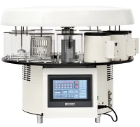



As a trusted supplier of slide stainers, I understand the paramount importance of staining quality control in the field of microscopy and pathology. Staining is a fundamental technique used to enhance the visibility of cellular and tissue structures, enabling accurate diagnosis and research. In this blog post, I will delve into the concept of staining quality control for slide stainers, exploring its significance, key components, and best practices.
Significance of Staining Quality Control
Staining quality control is essential for ensuring the reliability and reproducibility of staining results. In medical diagnostics, accurate staining is crucial for the detection and identification of diseases, such as cancer, infectious diseases, and autoimmune disorders. A poorly stained slide can lead to misinterpretation of results, potentially resulting in incorrect diagnoses and inappropriate treatment decisions.
In research settings, staining quality control is equally important for generating reliable data. Consistent staining results are necessary for comparing samples across different experiments and laboratories, ensuring the validity of research findings. Moreover, high-quality staining can enhance the visualization of cellular and molecular structures, facilitating a deeper understanding of biological processes.
Key Components of Staining Quality Control
1. Reagent Management
Reagents are the cornerstone of the staining process, and their quality and stability directly impact the staining results. As a slide stainer supplier, I emphasize the importance of using high-quality reagents from reputable manufacturers. Regularly check the expiration dates of reagents and discard any that are expired or show signs of degradation.
Proper storage of reagents is also crucial. Most staining reagents should be stored at specific temperatures and protected from light and moisture. Follow the manufacturer's instructions for storage conditions to maintain the integrity of the reagents.
2. Instrument Calibration and Maintenance
A well-calibrated and maintained slide stainer is essential for achieving consistent staining results. Regularly calibrate the instrument according to the manufacturer's recommendations to ensure accurate dispensing of reagents, proper timing of staining steps, and uniform staining across all slides.
Perform routine maintenance tasks, such as cleaning the staining chambers, replacing worn-out parts, and checking the fluid levels. A clean and well-maintained slide stainer reduces the risk of contamination and ensures the smooth operation of the instrument.
3. Slide Preparation
The quality of slide preparation can significantly affect the staining results. Ensure that the slides are clean and free of debris before applying the specimens. Use appropriate fixatives to preserve the cellular and tissue structures and prevent autolysis and putrefaction.
Properly section the specimens to achieve uniform thickness and avoid folding or wrinkling. The orientation of the specimens on the slides can also impact the staining results, so ensure that they are placed correctly.
4. Staining Protocol Optimization
Developing and optimizing a staining protocol is crucial for achieving the desired staining results. Consider factors such as the type of specimen, the staining method, and the specific reagents used. Conduct pilot experiments to determine the optimal staining conditions, including the concentration of reagents, the duration of staining steps, and the temperature.
Document the staining protocol in detail and follow it consistently to ensure reproducibility. Any changes to the protocol should be carefully evaluated and validated before implementation.
5. Quality Assurance and Quality Control Checks
Implement a comprehensive quality assurance (QA) and quality control (QC) program to monitor the staining process and ensure the reliability of the results. Regularly perform QC checks using control slides with known staining characteristics. Compare the staining results of the control slides with the expected results to identify any deviations or trends.
Establish acceptance criteria for the staining results and take corrective actions if the results fall outside the acceptable range. Keep detailed records of the QC checks, including the date, the type of control slide, the staining results, and any corrective actions taken.
Best Practices for Staining Quality Control
1. Training and Education
Provide comprehensive training to the users of the slide stainer on the proper operation of the instrument, the staining protocol, and the importance of staining quality control. Ensure that the users are familiar with the safety procedures and the proper handling of reagents.


Encourage continuous education and professional development to keep the users updated on the latest advancements in staining techniques and quality control practices.
2. Standardization
Standardize the staining process across different laboratories and operators to ensure consistency and comparability of the results. Use standardized staining protocols, reagents, and instruments to minimize variability.
Participate in external quality assessment programs to compare your staining results with those of other laboratories and identify areas for improvement.
3. Documentation and Record-Keeping
Maintain detailed records of all aspects of the staining process, including the reagent usage, the instrument calibration and maintenance, the slide preparation, and the staining results. These records are essential for traceability, quality control, and regulatory compliance.
Use a laboratory information management system (LIMS) to streamline the documentation process and facilitate data analysis.
4. Collaboration and Communication
Foster collaboration and communication among the laboratory staff, including the pathologists, technologists, and slide stainer operators. Encourage open dialogue and the sharing of knowledge and experiences to improve the staining quality control.
Establish a feedback mechanism to address any issues or concerns related to the staining process promptly.
Our Slide Stainers and Staining Quality Control
At our company, we are committed to providing high-quality slide stainers that are designed to meet the rigorous demands of staining quality control. Our Automated Stainer, Automatic Slide Stainer, and Automated Slide Stainer are equipped with advanced features and technologies to ensure accurate and consistent staining results.
Our slide stainers are designed for easy operation and maintenance, with user-friendly interfaces and intuitive controls. They are also compatible with a wide range of staining reagents and protocols, allowing for flexibility and customization.
In addition to providing high-quality slide stainers, we also offer comprehensive technical support and training services to our customers. Our team of experts is available to assist you with the installation, calibration, and maintenance of the slide stainer, as well as the optimization of the staining protocol.
Conclusion
Staining quality control is a critical aspect of the staining process, ensuring the reliability and reproducibility of staining results in medical diagnostics and research. By implementing a comprehensive quality control program that includes reagent management, instrument calibration and maintenance, slide preparation, staining protocol optimization, and quality assurance and quality control checks, you can achieve consistent and high-quality staining results.
As a slide stainer supplier, we are dedicated to helping our customers achieve the highest standards of staining quality control. If you are interested in learning more about our slide stainers or our staining quality control solutions, please contact us for a consultation. We look forward to working with you to meet your staining needs.
References
- Bancroft, J. D., & Gamble, M. (2008). Theory and Practice of Histological Techniques. Churchill Livingstone.
- Kiernan, J. A. (2008). Histological and Histochemical Methods: Theory and Practice. Oxford University Press.
- Luna, L. G. (1968). Manual of Histologic Staining Methods of the Armed Forces Institute of Pathology. McGraw-Hill.