


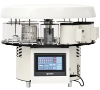


A tissue embedder is an essential instrument in histology laboratories, playing a crucial role in preparing tissue samples for microscopic examination. As a supplier of tissue embedders, I am well - versed in how these devices operate and their significance in the field of pathology. In this blog, I will take you through the detailed working mechanism of a tissue embedder.
The Basics of Tissue Embedding
Before delving into the working of a tissue embedder, it's important to understand the concept of tissue embedding. In histology, tissue samples need to be preserved and made firm enough to be cut into thin sections for microscopic analysis. This is achieved by embedding the tissue in a supportive medium, most commonly paraffin wax. The process of embedding encases the tissue within the wax, providing the necessary rigidity for sectioning with a microtome.
Components of a Tissue Embedder
A typical tissue embedder consists of several key components:
- Heating Chambers: These are used to melt the embedding medium, usually paraffin wax. The temperature in these chambers is precisely controlled to ensure that the wax remains in a liquid state at an appropriate viscosity for embedding.
- Cold Plate: The cold plate is a critical part of the embedder. After the tissue has been placed in the mold with molten wax, the mold is transferred to the cold plate. The cold plate rapidly cools the wax, solidifying it and embedding the tissue. You can find more information about a Tissue Embedding Center with cold plate.
- Work Surface: The work surface is where the actual embedding process takes place. It provides a stable area for handling the tissue samples, molds, and molten wax. It is often heated to prevent the wax from solidifying prematurely during the embedding process.
- Mold Holders: These are used to hold the embedding molds in place during the filling and cooling processes. They ensure that the molds are properly positioned and that the tissue remains in the desired orientation within the wax.
Step - by - Step Working Process
Step 1: Tissue Preparation
Before the tissue can be embedded, it must undergo a series of pre - embedding steps. First, the tissue is fixed to preserve its structure. This is typically done using formalin or other fixatives. After fixation, the tissue is dehydrated through a series of alcohol baths of increasing concentration to remove water from the tissue. This is followed by clearing, where the alcohol is replaced with a clearing agent such as xylene, which is miscible with both alcohol and paraffin wax.
Step 2: Wax Melting
The paraffin wax is placed in the heating chambers of the tissue embedder. The embedder is set to a specific temperature, usually around 56 - 60°C, depending on the type of wax being used. The heating elements in the chambers gradually heat the wax until it melts. The temperature is carefully regulated to maintain the wax in a liquid state with the right consistency for embedding.
Step 3: Tissue Placement in the Mold
Once the wax is molten, an appropriate embedding mold is selected. The size and shape of the mold depend on the size and type of the tissue sample. The tissue is carefully transferred from the clearing agent to the mold using forceps. It is important to position the tissue correctly within the mold to ensure that the desired sectioning plane will be obtained later.
Step 4: Filling the Mold with Wax
Using a pipette or a special pouring device, the molten wax is carefully poured into the mold until it completely covers the tissue. The wax fills all the spaces around the tissue, ensuring that it is fully encased. The work surface of the embedder is heated to prevent the wax from solidifying too quickly during this process.
Step 5: Cooling and Solidification
After the mold is filled with wax, it is immediately transferred to the cold plate. The cold plate is maintained at a low temperature, usually around 4 - 10°C. The rapid cooling causes the wax to solidify, embedding the tissue in a firm block. The cold plate ensures that the wax solidifies evenly, preventing the formation of air bubbles or uneven solidification that could affect the quality of the subsequent sections.
Step 6: Removal of the Embedded Block
Once the wax has completely solidified, the embedded block can be removed from the mold. This is usually a straightforward process, and the block is now ready for sectioning using a microtome. The embedded block provides the necessary support for cutting thin sections of the tissue, which can then be stained and examined under a microscope.
Advanced Features of Modern Tissue Embedders
Modern tissue embedders come with a range of advanced features that enhance their performance and usability. Some embedders are equipped with multiple heating chambers, allowing different types of wax or additives to be used simultaneously. This is useful for laboratories that work with a variety of tissue types or require different embedding protocols.


Many embedders also have programmable temperature controls. This allows the user to set specific temperatures for different stages of the embedding process, ensuring consistent results. Additionally, some embedders are designed with ergonomic features, such as adjustable work surfaces and easy - to - use controls, to reduce operator fatigue and improve efficiency.
You can explore more about these advanced features in a histology embedding station or a paraffin embedding station.
Importance of a Good Tissue Embedder
The quality of the tissue embedding process has a direct impact on the quality of the subsequent histological sections. A well - embedded tissue block will produce thin, uniform sections that are free from artifacts such as folds, tears, or air bubbles. This is essential for accurate diagnosis in pathology laboratories.
A reliable tissue embedder also improves the efficiency of the laboratory workflow. It reduces the time required for embedding, allowing more samples to be processed in a shorter period. This is particularly important in high - volume laboratories where a large number of tissue samples need to be analyzed on a daily basis.
Contact for Procurement
If you are in the market for a high - quality tissue embedder, we are here to help. Our tissue embedders are designed with the latest technology and advanced features to meet the diverse needs of histology laboratories. Whether you are a small research laboratory or a large - scale pathology center, we have the right solution for you. Contact us to discuss your requirements and start a procurement洽谈.
References
- Bancroft, J. D., & Gamble, M. (2008). Theory and Practice of Histological Techniques. Churchill Livingstone.
- Kiernan, J. A. (2008). Histological and Histochemical Methods: Theory and Practice. Oxford University Press.