


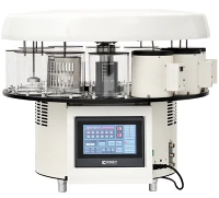
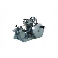


A microtome is an essential instrument in the fields of biology, medicine, and materials science, enabling researchers to obtain ultra-thin slices of specimens for microscopic examination. As a dedicated microtome supplier, I am thrilled to share insights into how these remarkable devices operate.
The Basic Principle of a Microtome
At its core, the function of a microtome is to cut extremely thin sections of a specimen. This is achieved through a precise mechanical or automated process that allows for controlled slicing. The basic principle involves advancing the specimen towards a sharp cutting edge, typically a blade, at a predetermined and adjustable thickness.
The key components of a microtome include the specimen holder, the blade holder, and the mechanism for advancing the specimen. The specimen is first embedded in a suitable medium, such as paraffin wax or resin, to provide support and maintain its structure during the cutting process. This embedded specimen is then securely placed in the specimen holder.
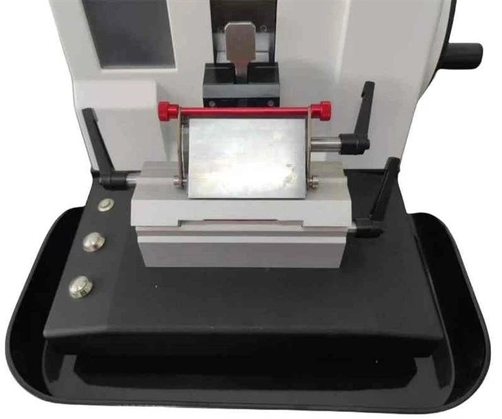
The blade holder holds the cutting blade in place at a precise angle relative to the specimen. The blade is usually made of high - quality materials such as stainless steel, glass, or diamond, depending on the nature of the specimen and the required section thickness. For example, glass blades are commonly used for cutting biological specimens embedded in paraffin wax, while diamond blades are preferred for harder materials or when extremely thin sections are needed.
Types of Microtomes and Their Working Mechanisms
Manual Microtome
A Manual Microtome is the most basic type of microtome. It is operated by hand, which gives the user a high degree of control over the cutting process. The user turns a handwheel to advance the specimen towards the blade. Each turn of the handwheel corresponds to a specific increment of specimen advancement, allowing for the adjustment of section thickness.
The working mechanism of a manual microtome involves a simple mechanical linkage. As the handwheel is turned, a screw - based or rack - and - pinion mechanism moves the specimen holder forward. The blade, held in a fixed position, then cuts through the advancing specimen, producing a thin section. Manual microtomes are often used in educational settings or for small - scale research where cost - effectiveness and simplicity are important.
Rotary Microtome
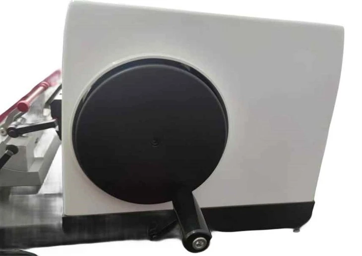
The Rotary Microtome is one of the most widely used types of microtomes. It operates on a rotary motion principle. A large handwheel is rotated, which drives a cam mechanism. This cam mechanism controls the up - and - down movement of the specimen holder as well as its forward advancement.
As the handwheel is turned, the specimen holder moves down towards the blade, and at the same time, it advances a small distance forward. When the specimen reaches the blade, a thin section is cut. The rotary motion allows for a smooth and continuous cutting process, which is especially useful for cutting large numbers of sections. Rotary microtomes can be adjusted to cut sections with a high degree of precision, typically ranging from a few micrometers to tens of micrometers in thickness.
Semi - automatic Microtome
A Semi - automatic Microtome combines the features of manual and fully automatic microtomes. It has an electric motor that can be used to drive the specimen advancement, while some functions, such as the up - and - down movement of the specimen holder during cutting, may still require manual operation.
The working mechanism of a semi - automatic microtome involves an electric motor connected to a gearbox. The motor rotates the gears, which in turn drive the specimen advancement mechanism. The user can set the desired section thickness using a control panel, and the motor will advance the specimen at the specified rate. This type of microtome offers a good balance between automation and user control, making it suitable for medium - scale research laboratories.
The Cutting Process
Regardless of the type of microtome, the cutting process generally follows a similar sequence. First, the specimen is properly aligned in the specimen holder to ensure that the desired area of the specimen is presented to the blade. The blade is then adjusted to the correct angle and position relative to the specimen.
Before starting the cutting, the section thickness is set according to the requirements of the experiment. For biological specimens, section thicknesses typically range from 3 to 10 micrometers for light microscopy, while thinner sections (less than 1 micrometer) may be required for electron microscopy.
As the microtome operates, the specimen is gradually advanced towards the blade. When the specimen makes contact with the blade, the sharp edge of the blade cuts through the specimen, separating a thin section. These sections are then collected on a glass slide or a suitable collecting medium for further processing, such as staining and mounting.
Factors Affecting the Cutting Quality
Several factors can affect the quality of the sections cut by a microtome. One of the most important factors is the sharpness of the blade. A dull blade can cause rough or uneven sections, tearing of the specimen, or compression artifacts. Regular blade sharpening or replacement is essential to maintain cutting quality.
The hardness and consistency of the specimen also play a crucial role. If the specimen is too hard, it may cause excessive wear on the blade or result in sections that are difficult to cut. On the other hand, if the specimen is too soft, it may deform during the cutting process. Proper embedding and fixation of the specimen can help to overcome these issues.
The cutting speed and the feed rate of the specimen also affect the cutting quality. A too - high cutting speed can cause the sections to be torn or damaged, while a too - low speed may result in uneven sections. The ideal cutting speed and feed rate depend on the type of specimen and the blade used.
Applications of Microtomes
Microtomes are used in a wide range of applications. In the field of biology, they are used to prepare tissue sections for histological examination. By cutting thin sections of tissues, researchers can study the cellular structure and organization, identify diseases, and understand biological processes.
In materials science, microtomes are used to prepare samples for microscopic analysis of materials such as metals, polymers, and ceramics. Thin sections of materials can reveal their internal structure, grain boundaries, and defects, which is important for understanding their properties and performance.
In the medical field, microtomes are used in pathology laboratories for the diagnosis of diseases. Pathologists examine thin sections of tissue samples to detect the presence of cancer cells, determine the stage of the disease, and guide treatment decisions.
Why Choose Our Microtomes
As a microtome supplier, we are committed to providing high - quality microtomes that meet the diverse needs of our customers. Our microtomes are designed with precision engineering, using the latest technology and high - quality materials.
We offer a wide range of microtomes, including manual, rotary, and semi - automatic models, to suit different budgets and research requirements. Our products are easy to operate, maintain, and provide consistent and high - quality cutting results.
Whether you are a researcher in a large - scale research institution, an educator in an academic setting, or a pathologist in a medical laboratory, our microtomes can help you achieve your research and diagnostic goals.
If you are interested in learning more about our microtomes or would like to discuss your specific needs, we encourage you to contact us for a detailed consultation. Our team of experts is ready to assist you in making the right choice for your laboratory.
References
- Humason, G. L. (1979). Animal Tissue Techniques. W. H. Freeman and Company.
- McManus, J. F. A., & Mowry, R. W. (1960). Histochemical and Cytochemical Methods. Little, Brown and Company.
- Stevens, A., & Lowe, J. S. (2000). Human Histology. Mosby.