


Can a cryotome be used for human tissue sectioning?
As a supplier of cryotomes, I am often asked whether our cryotomes can be used for human tissue sectioning. The answer is a resounding yes. Cryotomes are highly versatile instruments that have been widely employed in the field of histology for sectioning various types of tissues, including human tissues. In this blog post, I will delve into the details of how cryotomes work, their advantages for human tissue sectioning, and some key considerations when using them.
How Cryotomes Work
A cryotome, also known as a cryostat microtome, is a specialized instrument designed to cut thin sections of frozen tissues. The basic principle behind a cryotome involves rapidly freezing the tissue sample to a suitable temperature, typically between -20°C and -30°C, to make it firm enough for sectioning. Once the tissue is frozen, a sharp blade, usually a disposable microtome blade, is used to cut thin slices of the tissue. These slices can then be mounted on glass slides for further histological analysis, such as staining and microscopy.
The process of freezing the tissue is crucial as it helps to preserve the cellular structure and prevent the formation of ice crystals, which can damage the tissue and affect the quality of the sections. Most cryotomes are equipped with a refrigeration system that can maintain a constant low temperature inside the cutting chamber to ensure optimal freezing conditions.
Advantages of Using a Cryotome for Human Tissue Sectioning
- Rapid Sectioning: One of the main advantages of using a cryotome for human tissue sectioning is the speed at which sections can be obtained. Unlike traditional paraffin embedding methods, which can take several hours or even days to complete, cryotome sectioning can be done in a matter of minutes. This is particularly useful in clinical settings where rapid diagnosis is required, such as during intraoperative consultations.
- Preservation of Antigenicity: Cryotome sectioning also helps to preserve the antigenicity of the tissue, which is important for immunohistochemical staining and other molecular techniques. Since the tissue is not exposed to high temperatures or harsh chemicals during the freezing process, the antigens in the tissue remain intact and can be easily detected using specific antibodies.
- Flexibility: Cryotomes offer a high degree of flexibility in terms of section thickness and tissue type. They can be used to cut sections ranging from a few micrometers to several hundred micrometers thick, depending on the specific requirements of the experiment or analysis. Additionally, cryotomes can be used to section a wide variety of human tissues, including brain, liver, kidney, muscle, and skin.
- Real - Time Analysis: With cryotome sectioning, it is possible to perform real - time analysis of the tissue sections. This allows pathologists and researchers to quickly assess the quality of the sections and make any necessary adjustments to the cutting parameters. It also enables immediate feedback on the histological features of the tissue, which can be invaluable in clinical decision - making.
Key Considerations When Using a Cryotome for Human Tissue Sectioning
- Tissue Preparation: Proper tissue preparation is essential for obtaining high - quality cryotome sections. The tissue sample should be fresh and free from any contaminants. It is also important to ensure that the tissue is properly oriented and embedded in an appropriate embedding medium, such as OCT (optimal cutting temperature) compound, to facilitate sectioning.
- Blade Selection: The choice of blade is crucial for achieving smooth and clean sections. Disposable microtome blades are commonly used in cryotomes, and they come in different types and sizes. The blade should be sharp and free from any defects to minimize tissue damage and ensure accurate sectioning.
- Cutting Parameters: The cutting speed, temperature, and feed rate are important parameters that need to be optimized for each tissue type. These parameters can affect the quality of the sections, including their thickness,平整度, and integrity. It may take some trial and error to find the optimal cutting conditions for a particular tissue sample.
- Safety Precautions: When working with human tissues, it is important to follow all relevant safety precautions. This includes wearing appropriate personal protective equipment, such as gloves and lab coats, and ensuring that the cryotome is properly maintained and cleaned to prevent cross - contamination.
Our Cryotome Products
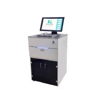
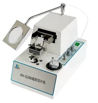
At our company, we offer a range of high - quality cryotomes that are suitable for human tissue sectioning. Our Cryotome models are designed with advanced features to ensure precise and efficient sectioning. For example, our Cryostats Semi - Automatic cryotomes offer semi - automated operation, which can save time and reduce the risk of operator error. Our Cryostat Microtome is a fully - featured instrument that provides excellent control over the cutting process and can produce high - quality sections consistently.
Conclusion
In conclusion, cryotomes are highly effective tools for human tissue sectioning. They offer numerous advantages, including rapid sectioning, preservation of antigenicity, flexibility, and real - time analysis. However, it is important to pay attention to key considerations such as tissue preparation, blade selection, cutting parameters, and safety precautions to ensure optimal results.


If you are interested in purchasing a cryotome for human tissue sectioning or have any questions about our products, we encourage you to contact us for a detailed discussion. Our team of experts is always ready to provide you with the best advice and support to meet your specific needs.
References
- Bancroft JD, Gamble M. Theory and Practice of Histological Techniques. 6th ed. Churchill Livingstone; 2008.
- Kiernan JA. Histological and Histochemical Methods: Theory and Practice. 4th ed. Springer; 2008.
- Mikel U, Giddings TH Jr. Biological Transmission Electron Microscopy: A Textbook. 2nd ed. Oxford University Press; 2007.