


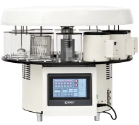
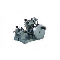


Hey there, fellow histology enthusiasts! Today, I'm super stoked to chat about one of the most essential tools in the histology field: the microtome. As a proud supplier of top - notch microtomes, I've seen firsthand how these amazing machines revolutionize the way we work in histology. So, let's dive right in and explore the various applications of a microtome in histology.
1. Sample Preparation for Microscopic Examination
The primary and most well - known application of a microtome in histology is sample preparation for microscopic examination. Before we can study the fine details of cells and tissues under a microscope, we need to cut them into extremely thin slices. And that's where the microtome comes in.
Tissues are usually first fixed to preserve their structure. Then, they're embedded in a suitable medium like paraffin wax or resin. Once embedded, the tissue block is mounted on the microtome. The microtome allows us to cut sections as thin as a few micrometers. These thin sections are crucial because they let light pass through, enabling us to clearly visualize the cellular and tissue architecture under a light microscope.
For example, in a pathology lab, when doctors are trying to diagnose a disease, they rely on high - quality tissue sections prepared by a microtome. A well - cut section can reveal abnormal cell growth, the presence of pathogens, and other important diagnostic features.
2. Research in Cell Biology
In the world of cell biology research, microtomes are indispensable. Scientists are constantly exploring the inner workings of cells, and microtomes help them get a closer look.


When studying the sub - cellular structures, such as organelles, we need to cut tissues in a way that preserves these delicate structures. Manual microtomes, like the ones you can find here, offer a high level of precision and control. Researchers can carefully adjust the cutting thickness and angle to obtain the best possible sections for their experiments.
For instance, if you're studying the mitochondria in muscle cells, you can use a microtome to cut thin sections of muscle tissue. These sections can then be stained and examined under an electron microscope to study the shape, size, and distribution of mitochondria within the cells.
3. Histological Teaching
Microtomes also play a vital role in histological teaching. In educational institutions, students need to learn how to prepare and examine tissue sections to understand the basic principles of histology.
Semi - automatic microtomes, like the ones available here, are great for teaching purposes. They offer a balance between manual control and automation. Students can learn the basic techniques of sectioning while having the assistance of some automated features.
For example, a semi - automatic microtome can have a preset cutting thickness, which helps students get consistent results. This way, students can focus on learning how to handle the tissue block, mount it correctly, and observe the sections under the microscope without getting overwhelmed by the technical details of cutting.
4. Industrial Quality Control
In industries related to pharmaceuticals, cosmetics, and food, microtomes are used for quality control. For example, in the pharmaceutical industry, when developing new drugs, it's important to study how the drug affects the tissues in the body.
Automatic microtomes, such as the ones you can check out here, are ideal for industrial applications. They can cut a large number of sections quickly and with high precision. This is crucial when you need to analyze a large sample size to ensure the safety and efficacy of a product.
In the food industry, microtomes can be used to examine the structure of food products. For example, when developing a new type of cheese, you can use a microtome to cut sections of the cheese to study its texture, the distribution of fat globules, and other quality - related features.
5. Forensic Histology
Forensic histology is another area where microtomes are extremely useful. In criminal investigations, pathologists may need to examine tissues from a crime scene to determine the cause of death or other important details.
Microtomes allow forensic pathologists to cut thin sections of tissues such as skin, muscle, and internal organs. These sections can then be stained and examined under a microscope to look for signs of injury, disease, or the presence of toxins.
For example, if a person has died under suspicious circumstances, a microtome - prepared tissue section can reveal if there are any signs of drug overdose, poisoning, or physical trauma at the cellular level.
Why Choose Our Microtomes?
As a microtome supplier, we understand the importance of having a reliable and high - quality microtome for your histology work. Our microtomes are designed with the latest technology and precision engineering. Whether you need a manual microtome for detailed research work, a semi - automatic microtome for teaching, or an automatic microtome for high - volume industrial applications, we've got you covered.
Our manual microtomes offer excellent control and precision, allowing you to cut the most delicate tissues with ease. The semi - automatic microtomes provide a user - friendly experience, making them perfect for beginners and educational settings. And our automatic microtomes are built for speed and efficiency, ensuring that you can get your work done quickly without compromising on quality.
If you're in the market for a new microtome or looking to upgrade your existing one, we'd love to have a chat with you. Contact us to discuss your specific needs and requirements. We can provide you with detailed product information, pricing, and even arrange a demonstration if you'd like. Let's work together to take your histology work to the next level!
References
- Kiernan, J. A. (2008). Histological and histochemical methods: theory and practice. Oxford University Press.
- Bancroft, J. D., & Gamble, M. (2008). Theory and practice of histological techniques. Churchill Livingstone.