




Enhancing staining details is a crucial aspect in various fields such as histology, cytology, and pathology. As a reputable Slide Stainer supplier, we understand the significance of achieving high - quality staining results. In this blog, we will explore several effective strategies to enhance the staining details with a Slide Stainer.
Understanding the Basics of Slide Staining
Before delving into the methods of enhancing staining details, it is essential to have a basic understanding of how slide staining works. Slide staining is a process of applying dyes or stains to biological specimens on a microscope slide to make the cells and tissues more visible under a microscope. Different stains are used depending on the type of specimen and the information that needs to be obtained.
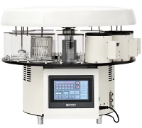
The Slide Stainer is a device that automates the staining process, ensuring consistency and reproducibility. It can handle multiple slides simultaneously, saving time and reducing the potential for human error. Our Automated Slide Stainer is designed with advanced technology to provide precise control over the staining process.
Optimizing Specimen Preparation
One of the first steps in enhancing staining details is to ensure proper specimen preparation. A well - prepared specimen is more likely to take up the stain evenly and clearly.
- Fixation: Fixation is the process of preserving the specimen to prevent decay and maintain its structure. The choice of fixative depends on the type of specimen and the stain that will be used. For example, formalin is a commonly used fixative for histological specimens. It is important to ensure that the fixation time is appropriate. Over - fixation can cause the specimen to become brittle and difficult to stain, while under - fixation may result in poor preservation and inconsistent staining.
- Dehydration: After fixation, the specimen needs to be dehydrated. This involves gradually replacing the water in the specimen with an organic solvent such as ethanol. Dehydration is crucial because most stains are dissolved in organic solvents. Incomplete dehydration can lead to poor stain penetration and uneven staining.
- Clearing: Clearing is the process of removing the dehydrating agent and replacing it with a clearing agent that is miscible with the embedding medium. Xylene is a commonly used clearing agent. Proper clearing ensures that the specimen is permeable to the embedding medium and the subsequent stains.
- Embedding: The specimen is then embedded in a solid medium such as paraffin wax. The embedding process should be carried out carefully to ensure that the specimen is oriented correctly and that the wax block is of good quality. A well - embedded specimen will provide thin and uniform sections, which are essential for high - quality staining.
Selecting the Right Stain
The choice of stain is another critical factor in enhancing staining details. Different stains have different affinities for various cellular components, and selecting the appropriate stain can highlight specific structures or molecules in the specimen.
- Hematoxylin and Eosin (H&E): H&E is one of the most commonly used stains in histology. Hematoxylin stains the nuclei blue - purple, while eosin stains the cytoplasm and extracellular matrix pink. This combination allows for clear visualization of the general structure of the tissue.
- Immunohistochemistry (IHC) Stains: IHC stains are used to detect specific proteins in the specimen. These stains use antibodies that bind to the target proteins, and a secondary detection system is used to visualize the bound antibodies. IHC stains can provide valuable information about the expression and localization of proteins in the cells and tissues.
- Special Stains: There are also many special stains available for specific purposes. For example, the Periodic Acid - Schiff (PAS) stain is used to detect carbohydrates, and the Masson's trichrome stain is used to differentiate between collagen, muscle, and other tissues.
Our Automated Stainer is compatible with a wide range of stains, allowing you to choose the most suitable stain for your specific application.
Adjusting the Staining Parameters
The Slide Stainer allows for precise control over several staining parameters, and adjusting these parameters can significantly enhance the staining details.
- Stain Concentration: The concentration of the stain can affect the intensity and clarity of the staining. A too - low concentration may result in weak staining, while a too - high concentration may cause over - staining and background noise. It is important to optimize the stain concentration based on the type of specimen and the stain used.
- Staining Time: The staining time is another important parameter. Different stains require different staining times to achieve optimal results. For example, some stains may need only a few minutes, while others may require several hours. Our Slide Stainer allows you to set the staining time accurately, ensuring consistent and reproducible staining results.
- Washing Steps: Washing steps are crucial to remove excess stain and prevent background staining. The number and duration of the washing steps should be optimized. Insufficient washing can lead to high background, while excessive washing may cause the stain to be washed out.
Maintaining the Slide Stainer
Regular maintenance of the Slide Stainer is essential to ensure its proper functioning and to obtain high - quality staining results.
- Cleaning: The Slide Stainer should be cleaned regularly to remove any residual stains, debris, or contaminants. This includes cleaning the staining chambers, the fluid delivery system, and the slide holders. Using appropriate cleaning agents and following the manufacturer's instructions is important to avoid damage to the instrument.
- Calibration: Calibration of the Slide Stainer is necessary to ensure accurate control of the staining parameters. This includes calibrating the fluid flow rate, the staining time, and the temperature. Regular calibration can help to maintain the consistency and reproducibility of the staining results.
- Reagent Management: Proper management of the staining reagents is also important. Reagents should be stored at the appropriate temperature and used within their expiration dates. Contaminated or expired reagents can lead to poor staining results.
Troubleshooting Common Staining Problems
Even with proper specimen preparation, stain selection, and parameter adjustment, staining problems may still occur. Here are some common staining problems and their solutions:
- Uneven Staining: Uneven staining can be caused by several factors, such as improper specimen preparation, inconsistent stain application, or problems with the Slide Stainer. To solve this problem, check the specimen preparation steps, ensure that the stain is applied evenly, and verify the proper functioning of the Slide Stainer.
- High Background Staining: High background staining can be due to insufficient washing, contaminated reagents, or over - staining. Increase the number and duration of the washing steps, replace the contaminated reagents, and adjust the stain concentration and staining time.
- Weak Staining: Weak staining may be caused by a low stain concentration, short staining time, or problems with the specimen. Increase the stain concentration, extend the staining time, and check the specimen preparation process.
Conclusion
Enhancing staining details with a Slide Stainer requires a combination of proper specimen preparation, appropriate stain selection, precise parameter adjustment, regular instrument maintenance, and effective troubleshooting. As a Slide Stainer supplier, we are committed to providing high - quality products and technical support to help you achieve the best staining results. Our Automated Stainer is designed with advanced features to meet the diverse needs of different applications.


If you are interested in improving your staining details and exploring our range of Slide Stainers, we invite you to contact us for procurement and further discussions. We look forward to working with you to enhance your staining processes and achieve outstanding results.
References
- Bancroft, J. D., & Gamble, M. (2008). Theory and Practice of Histological Techniques. Churchill Livingstone.
- Kiernan, J. A. (2008). Histological and Histochemical Methods: Theory and Practice. Oxford University Press.
- Cote, R. J., Pinkus, G. S., & Mills, S. E. (2013). Modern Surgical Pathology. Saunders.